Gallery
chondrocyte images: illustrations and micrographs. All credits and licenses listed.
Micrographs
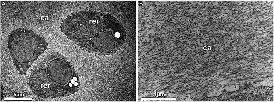
Hyaline cartilage TEM. Chondrocytes arranged in chondrons contain extensive rough endoplasmic reticulum (RER); surrounding matrix shows type II collagen fibrils interspersed with proteoglycans. Six-day-old mouse, demineralized bone.
Michael J.F. Blumer · CC BY 4.0 · source
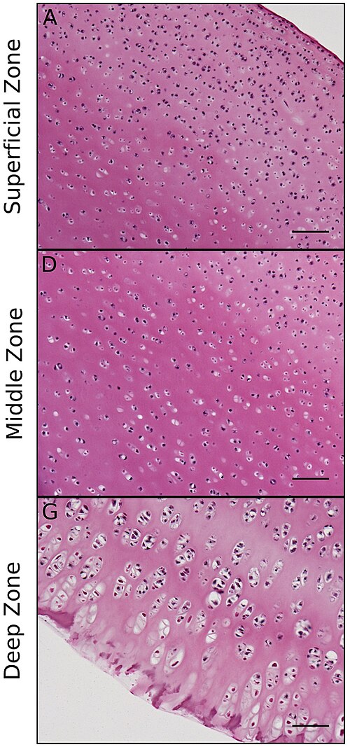
Articular cartilage ECM zonal architecture. Superficial zone: collagen II fibers aligned parallel to articular surface (resists shear). Deep zone: fibers aligned perpendicular to bone interface (absorbs compression).
Bautista, Park, Mazur, Aaron, Bilgen · CC0 · source
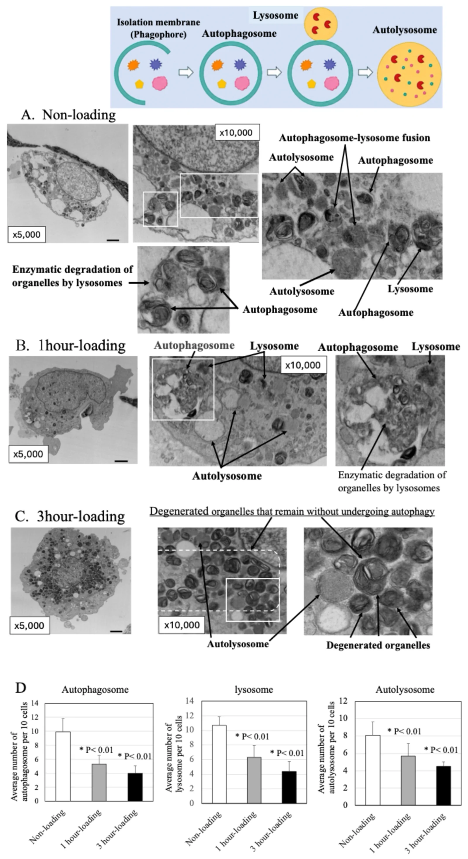
Human articular chondrocyte TEM (non-loading control arm). Panel A: autophagosomes, lysosomes, and autolysosomes showing normal autophagy activity. From a study of mechanical loading effects on OA chondrocytes; non-loading arm is the baseline control.
Sato, Iwata, Yamamoto et al 2026, Int J Mol Sci · CC BY 4.0 · source
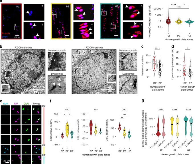
Human growth plate chondrocyte TEM. Resting zone (RZ) and proliferative zone (PZ) chondrocytes showing heterochromatin (asterisks) and lysosomes (arrowheads). Panel b from a study of cellular quiescence in the resting zone.
Avijgan, Lopez-Perez, Galicia et al 2026, Bone Research · CC BY 4.0 · source
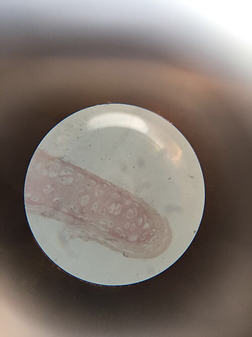
Hyaline cartilage histological section, H&E stain. Chondrocytes in lacunae visible within the cartilage matrix.
Nbnidhi · CC BY 4.0 · source
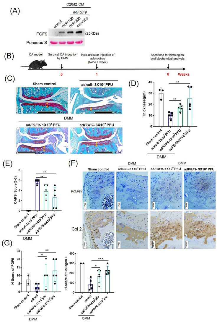
Safranin-O/Fast Green staining of knee articular cartilage. Red areas indicate preserved cartilage proteoglycans; green areas show depletion. From a DMM-induced OA model study of FGF9 intra-articular delivery.
Lo, Chen, Huang et al 2026, Bone and Joint Research · CC BY 4.0 · source
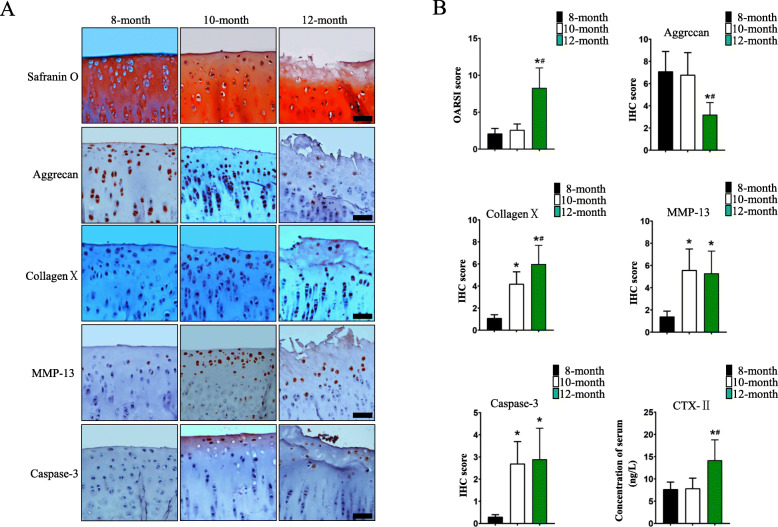
Tibial plateau articular cartilage, spontaneous OA. Safranin O and IHC for CollagenX, MMP-13, Aggrecan, Caspase-3 across age groups. Scale bar 100 um.
Wang, Wei, Xue et al 2021, BMC Musculoskeletal Disorders · CC BY 4.0 · source
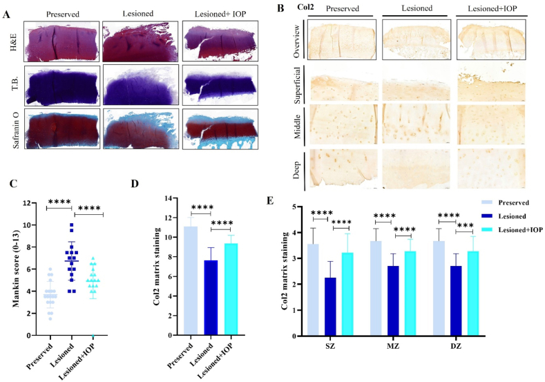
Human osteochondral explant H&E. Preserved (normal), lesioned (OA), and treated cartilage regions in the same section. Subchondral bone visible beneath cartilage.
Sayedipour, Mazzini, Tuerlings et al 2026, Osteoarthritis and Cartilage Open · CC BY 4.0 · source
Illustrations
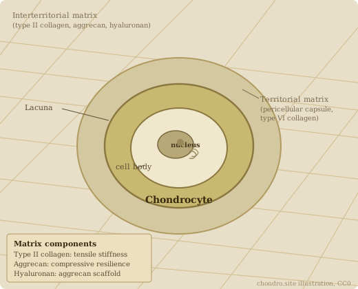
Chondrocyte in its lacuna. Territorial matrix (type VI collagen capsule) and interterritorial matrix (type II collagen, aggrecan, hyaluronan) shown.
Chondro / chondro.site · CC0
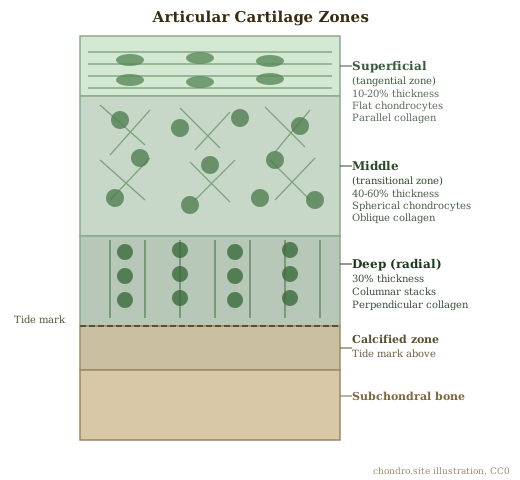
Articular cartilage zones: superficial (tangential), middle (transitional), deep (radial), calcified, and subchondral bone. Collagen fiber orientation shown per zone.
Chondro / chondro.site · CC0
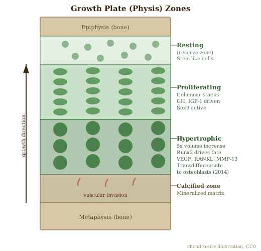
Growth plate zones: resting, proliferating, hypertrophic, calcified cartilage, and primary spongiosa.
Chondro / chondro.site · CC0