chondro.site

Everything there is to know about the chondrocyte, cited and in order.

Building the model…
Living model of the chondrocyte: the cell. Zoom between the stages. Morphology after Buckwalter JA, Mankin HJ, Instr Course Lect 1998 (PMID 9571449); Bhosale AM, Richardson JB, Br Med Bull 2008 (PMID 18676397).

Chondro. The chondrocyte has been studied since 1925. What do you need to know?

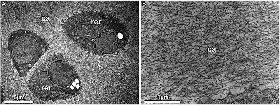
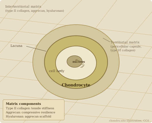
The chondrocyte is the only cell in cartilage. It lives alone in a lacuna, surrounded by matrix it made and maintains without blood supply or neighbouring contacts. It secretes type II collagen and aggrecan continuously, building the fibrillar meshwork and the proteoglycan aggregates that give cartilage its tensile strength and compressive resilience. In the growth plate it drives endochondral ossification: proliferating, hypertrophying, and signalling bone formation beneath it. In articular cartilage it is the whole maintenance crew for a tissue that must last a lifetime. When it fails, nothing replaces it. Source: Buckwalter and Mankin 1998, https://pubmed.ncbi.nlm.nih.gov/9571449/

2026-09-09 · PubMed
Chondrocyte inflammatory-immune responses in osteoarthritis: From pathogenesis to therapeutic intervention

Reviews how inflammatory and immune signaling drives cartilage degeneration in OA, beyond the usual biomechanical story.

2026-09-09 · PubMed
Cellular Senescence in Human Chondrocytes in Relation to Osteoarthritis

Reviews senescence as a hallmark linking chondrocyte aging to OA progression.

2026-09-09 · PubMed
Mitochondrial quality control modulating chondrocyte behavior and fate in knee OA

Covers mitophagy and mitochondrial dynamics as levers on chondrocyte survival and OA progression.

2026-09-09 · PubMed
Distinct adhesion energy and extracellular matrix profiles of healthy and osteoarthritic human chondrocytes

Biophysical comparison, healthy versus OA chondrocytes, adhesion energy and ECM differ measurably.

2026-09-09 · PubMed
Comparative Biophysical and Surface Property Analysis of Healthy and Osteoarthritic Chondrocytes

Surface property profiling relevant to cartilage tissue engineering, where autologous chondrocytes are the workhorse cell.

2026-09-09 · PubMed
Research progress of cuproptosis, ferroptosis, apoptosis, and autophagy in knee osteoarthritis

Copper and iron overload converge via cuproptosis, ferroptosis, apoptosis, and autophagy dysregulation to promote chondrocyte death and cartilage destruction.

2026-09-15 · Int Immunopharmacol
Selenoprotein S deficiency activates Wnt/beta-catenin signaling causing impaired terminal chondrocyte differentiation

Selenoprotein S (SelS) deficiency in chondrocytes activates Wnt/beta-catenin signaling and disrupts terminal differentiation; selenium deficiency linked to cartilage damage and osteoarticular disease; implicates SelS as a regulator of chondrocyte maturation via the Wnt pathway.

2026-09-09 · PMC
TGF-beta regulates phosphorylation and stabilization of Sox9 protein in chondrocytes through p38 and Smad dependent mechanisms

TGF-beta stabilizes Sox9 via p38 and Smad2/3; both pathways required independently. SOX9 protein level is a TGF-beta-dependent outcome.

2026-09-09 · PubMed
Sox9 family members negatively regulate maturation and calcification of chondrocytes through up-regulation of parathyroid hormone-related protein

Sox9 up-regulates PTHrP expression in association with Ihh/Gli2 signaling; the Ihh-PTHrP-Sox9 loop paces hypertrophic differentiation.

2026-09-09 · Frontiers
Molecular Mechanisms of Chondrocyte Proliferation and Differentiation

Comprehensive review: Sox9, Runx2, Ihh, PTHrP, TGF-beta, BMP, FGF, and Wnt signaling in chondrocyte fate decisions from mesenchymal condensation through hypertrophy.

2026-09-09 · PubMed
Transcriptional networks controlling chondrocyte proliferation and differentiation during endochondral ossification

Sox9 maintains chondrocyte fate; Runx2 drives hypertrophy; Ihh-PTHrP forms a feedback loop regulating the pace of hypertrophic differentiation.

2026-09-09 · PubMed
Expanded Stoichiometric Model of Chondrocyte Metabolism: Response to Cyclical Shear and Compressive Loading

Mechanotransduction modeled stoichiometrically under cyclical load; nitrogen availability is the primary constraint on matrix protein synthesis.

2026-09-09 · PubMed
Research progress on the mechanism of chondrocyte ferroptosis in osteoarthritis

Reviews iron-dependent lipid peroxidation, ferroptosis, as a cell death route in OA chondrocytes.

2026-09-09 · Springer
Identification of a putative progenitor-like chondrocyte subpopulation in osteoarthritic human cartilage

Identifies a chondrocyte subpopulation with progenitor-like markers inside OA cartilage, a possible repair reservoir.

2026-09-09 · Journal of Dental Research
Osteoarthritis and Chondrocytes: On the Road from Mechanisms to Treatment

Review tying chondrocyte mechanism work to where OA treatment is actually headed.

2026-09-09 · ScienceDirect
Age-associated regulation of chondrocyte hypertrophy in osteoarthritis: cartilage fate reprogramming

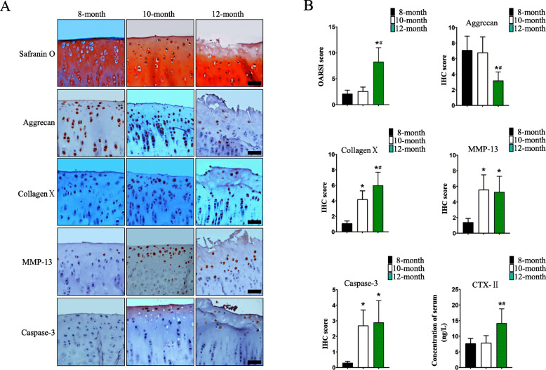
Aging lowers the threshold for the hypertrophy-like shift (collagen X, MMP13 up) via senescence, oxidative stress, mitochondrial dysfunction, and epigenetic drift.

2026-09-09 · PubMed
Advances in the mechanism and therapies of achondroplasia

FGFR3 gain-of-function mutation c.1138G>A (p.Gly380Arg) constitutively inhibits chondrocyte proliferation and differentiation; reviews targeted therapy landscape.

2026-09-09 · JCI
Fgfr3 enhancer deletion markedly improves all skeletal features in a mouse model of achondroplasia

CRISPR/Cas9 deletion of a cartilage-specific FGFR3 enhancer normalized long bone and vertebral growth and reduced foramen magnum stenosis in mice.

2026-09-09 · PMC
Advances and Challenges in the Pursuit of Disease-Modifying Osteoarthritis Drugs: A Review of 2010-2024 Clinical Trials

Reviews 11 DMOAD candidates including lorecivivint, sprifermin, UBX0101, TPX-100, GLPG1972, lutikizumab; none approved as of 2024.

2026-09-09 · Nature EMM
Disease-modifying therapeutic strategies in osteoarthritis: current status and future directions

Reviews anabolic, anti-catabolic, and anti-inflammatory DMOAD strategies; identifies cartilage matrix as the correct therapeutic target.

2026-09-09 · ScienceDirect
Update on clinical trials of intra-articular disease-modifying osteoarthritis drugs inducing cartilage regeneration

Intra-articular DMOAD trials 2025 update; sprifermin (FGF-18) showed cartilage thickness increase but not pain reduction; lorecivivint (Wnt/CLK2 inhibitor) in phase 3.

2026-09-09 · PubMed
Autologous chondrocyte implantation provides good long-term clinical results in the treatment of knee osteoarthritis: a systematic review

ACI showed sustained clinical improvement at long-term follow-up for focal chondral defects; MACI is the current US-approved product (Carticel phased out 2017).

2026-09-09 · PMC
Role and Effectiveness of Intra-articular Injection of Hyaluronic Acid in the Treatment of Knee Osteoarthritis: A Systematic Review

Hyaluronic acid injections improve pain and function in knee OA; effect size modest and duration variable; works best in early OA with preserved cartilage.

2026-09-09 · Mayo Clinic
Minimum 10-Year Outcomes of Matrix-Induced Autologous Chondrocyte Implantation in the Knee: A Systematic Review

MACI at minimum 10-year follow-up: significant durable improvement in patient-reported outcomes, satisfactory defect fill on MRI, low reoperation rate.

Cartilage-on-a-chip

All Cartilage-on-a-chip →
2026-09-10 · Cartilage
Exploring Osteoarthritis Dynamics: Patient-Specific Cartilage Samples in an Organ-on-a-Chip Model

Patient-derived OA cartilage held in an organ-on-a-chip to follow disease dynamics in human tissue.

2026-09-09 · PubMed
Exploring cartilage development and disease models: applications of cartilage organoids

Cartilage organoids as models for chondrogenesis and cartilage disease, covering differentiation, matrix synthesis, cultivation, and characterization.

2026-09-09 · PubMed
Growth plate organoid via layered induction in gelatin/alginate scaffold

Four-layer organoid recapitulates resting, proliferating, hypertrophic, and calcified zones of the native physis

2026-09-10 · Colloids Surf B Biointerfaces
GelMA hydrogel microsphere-based cartilage organoid precursors for the repair of cartilage defects

GelMA microspheres used to build cartilage organoid precursors, tested for defect repair.

2026-09-10 · Cell Stem Cell
Endothelialized callus organoids drive rapid regeneration of critical-size segmental long bone defects

Callus organoids carrying endothelium regenerate critical-size long bone defects; the cartilage callus route to bone, built in a dish.

2026-09-11 · ACS Biomater Sci Eng
Biomimetic COL-HA-PVA hydrogel with tuned pore architecture directs BMSC chondrogenesis for osteochondral regeneration

Hydrogel combining collagen, hyaluronic acid, and PVA with gradient porosity steers bone marrow stromal cells toward cartilage fate in osteochondral constructs.

2026-09-11 · Aesthetic Plast Surg
Comparative evaluation of exosomes, MSC, and nanofat on cartilage graft viability in rat model

Exosomes, MSC, and nanofat all improved graft viability; exosomes gave the best GAG and collagen II retention at 8 weeks. Thin; reconstructive model.

Spatial transcriptomics

All Spatial transcriptomics →
2026-09-09 · PubMed
CRIP1+ prehypertrophic chondrocytes: spatial transcriptomics confirms co-localization with prefibroblasts in OA

Spatial data places CRIP1+ preHTC at pathological niches alongside prefibroblasts; confirmed as diagnostic candidate

2026-09-09 · PubMed
OA single-cell and spatial transcriptomics: pathway regulation, cell interaction networks, therapeutic translation

August 2026 review integrating scRNA-seq and spatial data for OA; identifies actionable pathway targets

2026-09-10 · Int J Mol Med
Multi-omics integration in osteoarthritis: cell-type-specific gene-metabolite networks for precision medicine (Review)

Review joining transcriptomic and metabolomic layers by cell type in OA.

2026-09-09 · PubMed
The chondrocyte, architect of cartilage (Muir 1995)

Foundational framing: the chondrocyte as sole source and guardian of the cartilage matrix. Classic reference for matrix biology.

2026-09-09 · Royal Society B
From gristle to chondrocyte transplantation

History of cartilage repair and translational chondrocyte biology from the 1994 turning point onward.

2026-09-09 · PubMed
Articular cartilage: tissue design and chondrocyte-matrix interactions (Buckwalter 1998)

Classic review defining the three-component matrix (type II/IX/XI collagen meshwork, aggrecan-hyaluronan complexes, noncollagenous proteins) and their mechanical roles.

2026-09-09 · PubMed
Is cartilage matrix breakdown an appropriate therapeutic target in osteoarthritis? Aggrecan and collagen proteolysis

Reviews ADAMTS-mediated aggrecan cleavage and MMP-13-mediated collagen degradation as the two main matrix breakdown pathways in OA.
Substances tested 11

All substances →
Interleukin-1 alpha and interleukin-1 beta catabolic cytokine
acts on IL-1 receptor on the chondrocyte
Stimulate matrix proteoglycan degradation and inhibit glycosaminoglycan synthesis in cartilage explants. The reference catabolic insult: most chondroprotective compounds in this table are tested against it.
dose: not stated in the abstract  · bovine nasal cartilage explants; rabbit articular chondrocytes  · cartilage organ culture and chondrocyte culture  · Smith RJ, Chin JE, Sam LM, Justen JM 1991, Arthritis Rheum  · source
Interleukin-1 receptor antagonist protein (IRAP) receptor antagonist, the protein anakinra is built on
acts on IL-1 receptor, blocking IL-1 alpha and IL-1 beta binding
Concentration-dependent suppression of IL-1 effects in cartilage organ culture. Inhibited binding of radiolabelled IL-1 alpha to rabbit articular chondrocytes and suppressed collagenase, gelatinase, stromelysin and prostanoid production by IL-1-activated chondrocytes.
dose: 0.2 to 200 ng/ml, concentration-dependent  · bovine nasal cartilage explants; rabbit articular chondrocytes  · organ culture, receptor binding, enzyme assays  · Smith RJ, Chin JE, Sam LM, Justen JM 1991, Arthritis Rheum  · source
Engineered cationic IL-1 receptor antagonist protein therapeutic, charge-modified
acts on IL-1 receptor; the cationic charge drives uptake into the anionic cartilage matrix
Outperforms anakinra at joint retention and at preventing IL-1-induced cartilage inflammation. The problem it solves is residence time: anakinra clears the joint faster than the inflammation resolves.
dose: not stated in the abstract  · not stated in the abstract  · joint retention and cartilage inflammation assays  · Boyer TL et al. 2026, Osteoarthritis Cartilage  · source
Lorecivivint (SM04690) small-molecule intranuclear kinase inhibitor, Wnt pathway
acts on CDC-like kinase 2 (CLK2) and dual-specificity tyrosine phosphorylation-regulated kinase 1A (DYRK1A)
Inhibits CLK2-mediated phosphorylation of SR splicing factors and DYRK1A-mediated phosphorylation of SIRT1 and FOXO1, modulating Wnt without touching beta-catenin. CLK2 inhibition induces early chondrogenesis; DYRK1A inhibition enhances mature chondrocyte function and carries the anti-inflammatory effect through NF-kB and STAT3. In the rat MIA model a single intra-articular injection increased joint cartilage, decreased pain and improved weight-bearing.
dose: single intra-articular injection, dose not stated in the abstract  · human mesenchymal stem cells, chondrocytes and synovial fibroblasts in vitro; rat monosodium iodoacetate OA model  · kinase assays, western blot, siRNA knockdown, qPCR, in vivo OA model  · Deshmukh V et al. 2019, Osteoarthritis Cartilage  · source
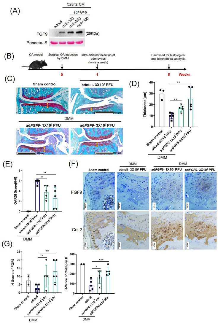
Sprifermin (recombinant human FGF-18) growth factor, disease-modifying OA drug candidate
acts on FGF receptor signalling in the chondrocyte
Promotes chondrocyte proliferation and extracellular matrix synthesis, increases cartilage thickness in a dose-dependent manner, and inhibits proteolytic enzyme activity. Reviewed as an effective DMOAD acting by both anti-catabolic and excitoanabolic routes.
dose: dose-dependent thickness increase reported; the doses are not stated in the abstract  · review across models  · review  · Song Z et al. 2021, Front Cell Dev Biol  · source
Sprifermin in explant storage medium growth factor, tissue preservation use
acts on FGF receptor signalling during cold storage of osteochondral allograft tissue
Tested for whether adding sprifermin during storage preserves the depth-dependent mechanical inhomogeneity of articular cartilage, the property fresh allografts lose at 4 degrees C.
dose: not stated in the abstract  · articular cartilage explants, species not stated in the abstract  · explant storage and depth-dependent mechanical testing  · Meloni GR et al. 2019, Eur Cell Mater  · source

Public datasets 12

GSE114007 RNA-seq, 38 samples Homo sapiens
Identification of transcription factors responsible for dysregulated networks in human osteoarthritis cartilage by global gene expression analysis
Fisch KM et al. 2018, Osteoarthritis Cartilage  · paper  · public at NCBI GEO; cited in 48 abstracts of the sweep, the most reused chondrocyte dataset found  · accession resolved 2026-09-12 via eutils esummary
GSE57218 expression microarray, 73 samples Homo sapiens
Gene expression profiles from joint-matched macroscopically intact and OA affected cartilage of patients undergoing joint replacement surgery
Ramos YF et al. 2014, PLoS One  · paper  · public at NCBI GEO; joint-matched intact against affected cartilage from the same patient, which controls for donor  · accession resolved 2026-09-12 via eutils esummary
GSE55235 expression microarray, 30 samples Homo sapiens
Identification of rheumatoid arthritis and osteoarthritis patients by transcriptome-based rule set generation
Woetzel D et al. 2014, Arthritis Res Ther  · paper  · public at NCBI GEO; cited in 35 abstracts of the sweep  · accession resolved 2026-09-12 via eutils esummary
GSE55457 expression microarray, 33 samples Homo sapiens
Identification of rheumatoid arthritis and osteoarthritis patients by transcriptome-based rule set generation
Woetzel D et al. 2014, Arthritis Res Ther  · paper  · public at NCBI GEO; companion series to GSE55235 from the same study  · accession resolved 2026-09-12 via eutils esummary
GSE51588 expression microarray, 50 samples Homo sapiens
Genome-wide Expression Profiles of Subchondral Bone in Osteoarthritis
Chou CH et al. 2013, Arthritis Res Ther  · paper  · public at NCBI GEO; subchondral bone rather than cartilage, the tissue on the other side of the tidemark  · accession resolved 2026-09-12 via eutils esummary
GSE82107 expression microarray, 17 samples Homo sapiens
Synovial biopsies of osteoarthritis patients
Broeren MG et al. 2016, PLoS One  · paper  · public at NCBI GEO; synovium, not cartilage, and included because the sweep shows it paired with cartilage sets  · accession resolved 2026-09-12 via eutils esummary
GSE169077 expression microarray, 11 samples Homo sapiens
Dysregulated gene expression in human osteoarthritis cartilage
no linked publication listed in GEO at the date resolved  · public at NCBI GEO; cited in 25 abstracts of the sweep despite carrying no linked paper  · accession resolved 2026-09-12 via eutils esummary
GSE117999 expression microarray, 24 samples Homo sapiens
Transcriptome comparison of cartilage from patients with and without osteoarthritis
no linked publication listed in GEO at the date resolved  · public at NCBI GEO  · accession resolved 2026-09-12 via eutils esummary
GSE324993 single-cell RNA-seq Homo sapiens
Single-cell RNA sequencing of chondrocytes from osteoarthritis (OA) patients and healthy controls
no linked publication listed in GEO at the date resolved  · public at NCBI GEO; single-cell resolution, which the older bulk sets above do not give  · accession resolved 2026-09-12 via eutils esummary
GSE308009 single-cell RNA-seq Mus musculus
Inhibition of the Gerozyme 15-Prostaglandin Dehydrogenase Promotes Articular Cartilage Regeneration in Osteoarthritis due to Injury or Aging [scRNA-Seq]
Singla M et al. 2026, Science  · paper  · public at NCBI GEO  · accession resolved 2026-09-12 via eutils esummary
GSE232109 RNA-seq, high throughput sequencing Homo sapiens
Recapitulation of endochondral ossification by human pluripotent stem cell-derived SOX9+ sclerotomal progenitors
Xiong J et al. 2025, Nat Commun  · paper  · public at NCBI GEO  · accession resolved 2026-09-12 via eutils esummary
GSE296290 single-cell multiome Bos taurus
Leveraging single cell multiomic analyses to identify gene regulatory networks that drive human articular cartilage cell fate
Jachim SK et al. 2026, Osteoarthritis Cartilage  · paper  · public at NCBI GEO; GEO lists the organism as Bos taurus while the series title says human, so read the series page before reuse  · accession resolved 2026-09-12 via eutils esummary

Digital twin

Open the twin →
13 um acrossOne cell at a fixed scale; the circle grows and shrinks with the slider.
Matrix held per unit of cell 88.8 fold 24.4 to 195
Turnover in units of the cell 0.648 cell volumes/day 0.178 to 1.42
Open the twin →

Chondro's summary

Everything here waited its turn. Nothing here is rushed and nothing here is unsourced.


History of discovery

Year / period 1925 · Event Benninghoff described articular cartilage zones by collagen fiber orientation and chondrocyte distribution using light microscopy. The arcade model of collagen organization dates to this work.
Year / period 1934-1939 · Event Hyaluronic acid named (1934) and demonstrated in cartilage (1939). Matrix composition begins to be traceable.
Year / period Mid-20th century · Event Isotopic tracer methods (35S) clarified chondrocyte metabolic roles. The cell was no longer inert filler.
Year / period 1995 · Event Muir's "The chondrocyte, architect of cartilage" defines the cell as the sole source and guardian of the cartilage matrix. A foundational framing.
Year / period 2014 · Event Three independent groups demonstrated by in vivo lineage tracing that hypertrophic chondrocytes transdifferentiate into osteoblasts, overturning 40 years of apoptosis dogma.

Function

Claim Sole cell in cartilage · Detail The chondrocyte is the only cell in hyaline and articular cartilage; no vasculature, nerves, or lymphatics enter the tissue. All matrix maintenance falls to this cell alone.
Claim Zone-specific behavior · Detail Superficial zone chondrocytes carry a distinct catabolic and senescent signature versus deeper zones, per single-cell RNA-seq of 72,616 cells from 11 human samples.
Claim Biophysical distinction · Detail Healthy and osteoarthritic chondrocytes differ measurably in adhesion energy and ECM profile.
Claim Zone-differential load response · Detail Chondrocytes from superficial versus deep zones respond differently to oscillatory tensile loading; functional specialization is zone-intrinsic.
Claim Compression sensing · Detail Compressive stress drives defined mechanosignaling pathways in chondrocytes, reviewed toward scaffold engineering.
Claim Nitrogen limits matrix synthesis · Detail Stoichiometric modeling under cyclical load: nitrogen availability is the primary constraint on chondrocyte matrix protein synthesis.

Cartilage types

Type Hyaline · Matrix composition Glassy, homogeneous; high aggrecan and bound water · Collagen Type II, IX, XI · Location Articular surfaces, trachea, costal, nasal, fetal skeleton
Type Fibrocartilage · Matrix composition Dense fibrous; high collagen-to-proteoglycan ratio · Collagen Type I and II · Location Knee meniscus, intervertebral discs, pubic symphysis, tendon insertions
Type Elastic · Matrix composition Dense elastic fiber network plus collagen · Collagen Type II plus elastin · Location Epiglottis, pinna, auditory tube, laryngeal cartilages

No primary citation for the three-type classification itself; secondary structural sources cited above.


Structure: articular cartilage zones

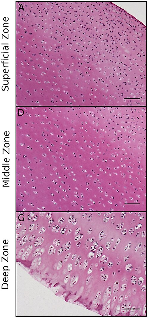
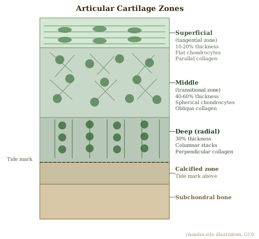
Four zones from surface to bone. Zone identity is determined by collagen orientation, chondrocyte shape, and proteoglycan density.

Zone Superficial (tangential) · Depth (approx) 10-20% of thickness · Collagen orientation Parallel to articular surface · Chondrocyte morphology Flattened, disk-shaped
Zone Middle (transitional) · Depth (approx) 40-60% of thickness · Collagen orientation Oblique, no preferred direction · Chondrocyte morphology Spherical, clustered
Zone Deep (radial) · Depth (approx) 30% of thickness · Collagen orientation Perpendicular to articular surface · Chondrocyte morphology Spherical, columnar
Zone Calcified · Depth (approx) Below tide mark · Collagen orientation Perpendicular; calcified matrix · Chondrocyte morphology Hypertrophic-like, embedded

Benninghoff's 1925 arcade model: collagen fibers arch from deep perpendicular bundles through the middle zone to the tangential superficial layer, creating a self-bracing architecture.


Matrix biology

The cartilage matrix is the product of chondrocyte synthesis and the medium through which the chondrocyte senses mechanical load.

Component Type II collagen · Class Fibrillar collagen · Function Tensile stiffness and strength; forms the fibrillar meshwork
Component Type IX collagen · Class FACIT collagen · Function Cross-links type II fibrils; surface molecule on fibrils
Component Type XI collagen · Class Fibrillar collagen · Function Regulates fibril diameter; found within the fibril alongside type II
Component Aggrecan · Class Large aggregating proteoglycan · Function Compressive stiffness and resilience; binds water via sulfated glycosaminoglycans
Component Hyaluronan · Class Glycosaminoglycan · Function Backbone for aggrecan aggregates; space-filling; non-sulfated
Component Noncollagenous proteins · Class Mixed class · Function Cell attachment, matrix organization, signaling (fibronectin, matrilin, COMP)
Component ADAMTS-4/5 · Class Aggrecanase · Function Principal aggrecan-cleaving enzymes in OA; therapeutic target candidate
Component MMP-13 · Class Collagenase · Function Dominant collagenase in OA cartilage breakdown

Development: growth plate and endochondral ossification

Most bones form through endochondral ossification. The growth plate is the engine.

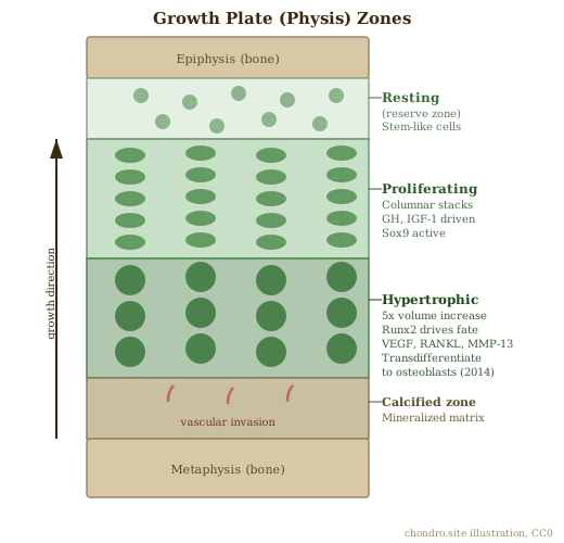
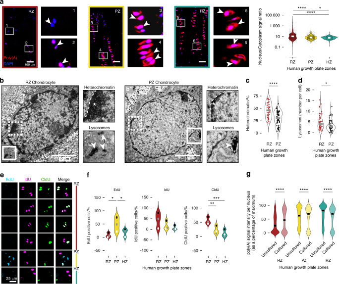
Growth plate zone Resting (reserve) · Cell state Quiescent chondrocytes · Activity Stem-like; source of proliferating cells
Growth plate zone Proliferating · Cell state Rapidly dividing chondrocytes · Activity Columnar stacks; driven by growth hormone and IGF-1
Growth plate zone Hypertrophic · Cell state Enlarged chondrocytes · Activity Volume increase; mineralizes matrix; produces VEGF, RANKL, MMP-9/13
Growth plate zone Calcified cartilage · Cell state Terminally differentiated · Activity Matrix mineralized; invaded by blood vessels and osteoblasts

Sox9 maintains chondrocyte fate. Runx2 drives hypertrophy. Indian hedgehog (Ihh) and parathyroid hormone-related peptide (PTHrP) form a feedback loop regulating the pace of hypertrophic differentiation. Source: Wuelling 2010.

Claim Hypertrophic chondrocytes transdifferentiate · Detail In vivo lineage tracing: hypertrophic chondrocytes give rise to osteoblasts in development, postnatal growth, and fracture healing. 2014 revision overturns 40 years of apoptosis dogma.
Claim Hypertrophic chondrocytes as stem cell reservoir · Detail Hypertrophic chondrocytes serve as a reservoir for marrow-associated skeletal stem and progenitor cells, osteoblasts, and adipocytes during skeletal development.
Claim Hypertrophy regulatory significance · Detail Precise regulation of chondrocyte hypertrophy is essential for homeostasis and repair, not only for pathological outcomes.
Claim Cartilage organoids · Detail Organoids now model chondrogenesis and cartilage disease with architecture, cellular heterogeneity, and ECM properties close to native tissue.
Claim Resting zone stem-like cells · Detail The human growth plate resting zone holds subpopulations with features of quiescent stem cells.
Claim Clinical regeneration · Detail Matrix-associated chondrocyte implantation, scaffolds, and controlled-release biologics are advancing toward mechanism-based cartilage repair.
Claim Glucocorticoid-induced ferroptosis in growth plate · Detail Dexamethasone silences lncRNA MALAT1 by DNA methylation, unleashing miR-124-3p to repress LPCAT3 and trigger ferroptosis in growth plate chondrocytes.

Pathology: osteoarthritis

Claim Inflammatory-immune axis · Detail OA progression is tied to inflammatory and immune signaling in chondrocytes, not just biomechanics and matrix breakdown.
Claim Hypertrophy as drug target · Detail Age-associated chondrocyte hypertrophy is a tractable node for disease-modifying OA drugs; driven by senescence, oxidative stress, and epigenetic drift.
Claim Senescence · Detail Chondrocyte cellular senescence is linked to OA as an aging hallmark.
Claim Mitochondrial quality control · Detail Mitophagy and mitochondrial dynamics shape chondrocyte fate in knee OA.
Claim Ferroptosis · Detail Iron-dependent lipid peroxidation is a chondrocyte death route in OA.
Claim Cuproptosis · Detail Copper overload activates cuproptosis alongside ferroptosis, apoptosis, and autophagy dysregulation; convergent cell death drives cartilage destruction.
Claim Oxiapoptophagy · Detail IL-1beta drives simultaneous oxidative stress, apoptosis, and autophagy in chondrocytes.
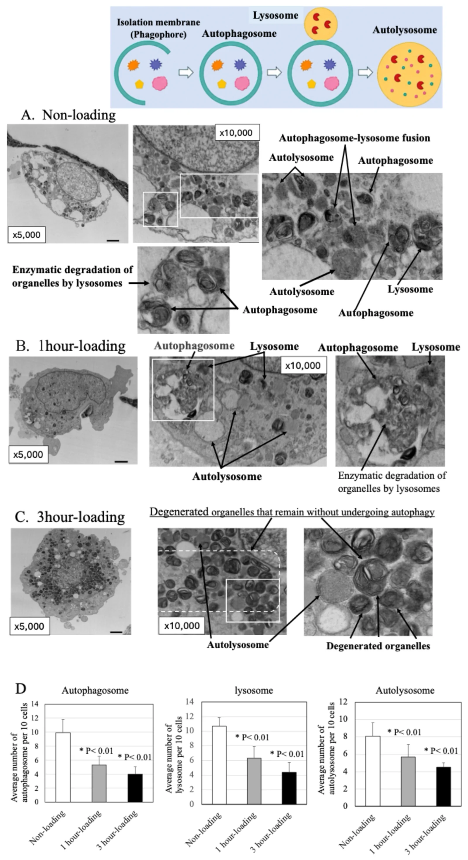
Claim Autophagy dual role · Detail Normal autophagy is chondrocyte-protective; its decline permits damaged organelle accumulation and ECM degradation.
Claim Programmed cell death landscape · Detail Seven distinct programmed death pathways regulate chondrocyte fate through interlocking molecular networks in OA.
Claim Progenitor-like reservoir · Detail A progenitor-like chondrocyte subpopulation with distinct markers sits inside OA cartilage, a possible repair reservoir.
Claim Epigenetic brake on matrix loss · Detail DOT1L-mediated H3K79me3 of ITCH promotes AURKA ubiquitination and suppresses ECM degradation in OA.
Claim Ferroptosis as a drug target · Detail Vestitol reduces ferroptosis-associated features and OA progression through GSK3B/Nrf2/GPX4. Preclinical.
Claim Senescence-targeted therapy · Detail Senolytic and senomorphic drug candidates against senescent chondrocytes are under review as OA interventions.
Claim m6A-mediated ferroptosis · Detail KIAA1429 suppresses USP3 via m6A methylation, accelerating chondrocyte senescence and ferroptotic death in OA.
Claim Local vs systemic OA · Detail OA has both local mechanical and systemic metabolic/inflammatory drivers; the two are not separable in late disease.
Claim Lactylation reprograms chondrocyte fate · Detail Lactate accumulation in the OA joint drives protein lactylation that shifts chondrocytes toward catabolic and senescent states and sustains joint inflammation; a metabolic axis on top of the cytokine story.
Claim Mitochondrial homeostasis as unifying target · Detail Mitochondrial dysfunction is a convergent pathogenic mechanism across cartilage, bone, and muscle degeneration; quality control pathways are precision targets for chondrocytes.
Claim TMJ OA: lncRNA-OIP5-AS1 axis · Detail lncRNA OIP5-AS1 sequesters miR-223-3p to relieve FoxO3 repression and protect chondrocytes from apoptosis in temporomandibular joint OA; extends the lncRNA-sponge pattern to a non-knee joint.

Medications

Substances tested on the chondrocyte, cited and linked. Dose and species on every row.

Substance IL-1Ra (IRAP) · Class Cytokine antagonist · Effect Blocks IL-1 receptor; reduces matrix degradation · Species/model Human synovial cells
Substance Lorecivivint · Class CLK/DYRK inhibitor · Effect Inhibits Wnt and inflammatory pathways; phase II for knee OA · Species/model Human (clinical)
Substance Sprifermin (FGF18) · Class Recombinant growth factor · Effect Anabolic: stimulates chondrocyte proliferation and cartilage thickness; phase II · Species/model Human (clinical)
Substance Dexamethasone · Class Glucocorticoid · Effect Anti-inflammatory at short course; drives growth plate chondrocyte ferroptosis via MALAT1 methylation at sustained doses · Species/model Rat (growth plate)
Substance Eugenol · Class Natural phenylpropanoid · Effect Sustains ECM homeostasis by balancing ALK1 and ALK5 arms of TGF-beta signaling · Species/model Chondrocyte cell line
Substance Secoisolariciresinol diglucoside · Class Plant lignan · Effect Reduces IL-1beta-driven inflammation through ERBB2-coupled JAK2/STAT3 suppression · Species/model OA chondrocyte model
Substance Chlorogenic acid · Class Dietary polyphenol · Effect Activates Nrf2/HO-1, suppresses ferroptotic cartilage loss in preclinical OA · Species/model Rodent OA model
Substance Vestitol · Class Phytoalexin isoflavonoid · Effect Reduces ferroptosis features and OA progression through GSK3B/Nrf2/GPX4. Preclinical. · Species/model Rodent OA model
Substance Metformin · Class Biguanide, AMPK activator · Effect Chondroprotective through AMPK/SIRT1-driven autophagy and NF-kB suppression; review of OA applications. · Species/model Review (multiple models)
Substance Salvianolic acid B (nano-assembly) · Class Phenolic acid (Traditional Chinese Medicine) · Effect Restores mitochondrial function in chondrocytes via PINK1/Parkin-mediated mitophagy; preclinical OA · Species/model Rodent OA model
Substance Epimedium polysaccharides (fermented) · Class Herbal polysaccharide · Effect Gut microbial fermentation into SCFAs substantially enhances chondroprotective activity; gut-joint axis · Species/model Rodent model

Cartilage-on-a-chip

Thin. One primary study this pass. The idea is sound; the record is short.

Claim Patient tissue on a chip · Detail Patient-specific osteoarthritic cartilage samples have been maintained in an organ-on-a-chip model to study disease dynamics.

Organoids

Claim Cartilage organoids as models · Detail Organoids now model chondrogenesis and cartilage disease with architecture, cellular heterogeneity, and ECM properties close to native tissue.
Claim Growth plate organoid · Detail A layered organoid in a gelatin/alginate scaffold reproduces the growth plate zone sequence.
Claim Organoid precursors for repair · Detail GelMA hydrogel microspheres serve as cartilage organoid precursors for defect repair.
Claim Callus organoids · Detail Endothelialized callus organoids regenerate critical-size segmental long bone defects.
Claim Biomimetic hydrogel chondrogenesis · Detail A COL-HA-PVA hydrogel with gradient porosity steers bone marrow stromal cells toward cartilage fate in osteochondral constructs.
Claim Exosomes outperform MSC for graft viability · Detail Exosomes, MSC, and nanofat all improved cartilage graft viability in a rat model; exosomes gave best GAG and collagen II retention at 8 weeks. Thin; reconstructive model.

Spatial transcriptomics

Claim CRIP1+ prehypertrophic hub · Detail Spatial data place CRIP1+ prehypertrophic chondrocytes beside prefibroblasts in OA cartilage.
Claim Review of the field · Detail Single-cell and spatial transcriptomics of OA: pathway regulation, cell interaction networks, therapeutic translation.
Claim Multi-omics by cell type · Detail Gene and metabolite networks resolved by cell type in OA, reviewed for precision medicine.

Decalcification

Time and concentration are primary variables. Cited or marked lab-specific.

Agent EDTA (chelating) · Concentration ~10% (lab-specific, verify) · Time 1-3 weeks at room temp; 3-7 days at 37 C (lab-specific, verify) · Endpoint / notes Preferred for morphology and antigen preservation; confirm with X-ray and needle probe · IHC and molecular impact Best for IHC, in situ hybridization, molecular assays; HIER works normally
Agent Formic acid, short course · Concentration Lab-specific · Time Under 5 cycles of 6 hours each · Endpoint / notes Acceptable; check endpoint at 24 h and 48 h · IHC and molecular impact Does not alter antigenicity; allows mutation, amplification, fusion transcript detection
Agent Formic acid, long course · Concentration Lab-specific · Time More than 5 cycles of 6 hours · Endpoint / notes Over-decal risk; avoid when IHC planned · IHC and molecular impact False-negative IHC and molecular results
Agent HCl · Concentration Lab-specific · Time Lab-specific · Endpoint / notes Avoid for downstream molecular work · IHC and molecular impact False-negative IHC and molecular analysis
Agent EDTA, accelerated (hypertonic saline + detergent) · Concentration Not specified · Time Shorter than standard EDTA · Endpoint / notes Speeds decal without acid tradeoff · IHC and molecular impact Preserves mRNA better than standard EDTA
Agent Nitric acid, 0.1% (ovine osteochondral) · Concentration 0.1% · Time 48 hours · Endpoint / notes Best speed-preservation balance in this model · IHC and molecular impact Not assessed for IHC in source
Agent EDTA + nitric acid, combined · Concentration Not specified · Time Faster than EDTA alone · Endpoint / notes Rapid substantial tissue degradation; avoid · IHC and molecular impact Reduced histological quality

Nothing good happens fast here. Acid buys speed and spends it in false negatives. Sourced three times now across two tissue systems. The pattern holds.


Methods and stains

Thin: no stain-specific primary citations yet beyond decal-IHC compatibility (see Decalcification). Sources remain structural histology texts.

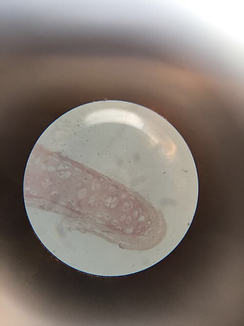
Method H and E · Use Routine morphology; chondrocyte nuclei, lacunae, matrix basophilia · Notes Basophilia of cartilage matrix reflects proteoglycan content
Method Safranin O / Fast Green · Use Proteoglycan distribution in cartilage · Notes Loss of Safranin O staining is a hallmark of OA-related aggrecan depletion
Method Alcian blue · Use Glycosaminoglycan content, particularly in growth plate · Notes pH-dependent; 0.1 M HCl distinguishes sulfated from non-sulfated
Method Masson's trichrome · Use Collagen fibers in fibrocartilage and bone · Notes Distinguishes cartilage from fibrous tissue at insertion zones
Method Type II collagen IHC · Use Confirms hyaline vs fibrocartilage identity · Notes EDTA decal required; acid decal may compromise the signal

Quality and regulation

Compliance claims wait for a standard, not a vendor page.

Claim CAP pre-analytical reporting · Detail CAP reporting templates require pre-analytical variables including decalcification to be reported where they may affect test results.
Claim Lab validation required · Detail ASCO-CAP guidance (HER2 update) requires labs to run their own validation studies for a test method rather than trust published protocols; the same logic applies to decal-affected IHC.
Claim Endpoint testing, general · Detail Vendor orientation material covers decal chemistry and endpoint testing at a general level; not lab-validated data; marked thin until a primary or standards source confirms.

CLIA-specific decal QC requirement: not found across four search passes. Section stays thin and open. No ASCP position statement on decalcification located. Both remain active open searches, not filled with a guess.

Latest 8

2026-09-15 · Int Immunopharmacol
Selenoprotein S deficiency activates Wnt/beta-catenin signaling causing impaired terminal chondrocyte differentiation

Selenoprotein S (SelS) deficiency in chondrocytes activates Wnt/beta-catenin signaling and disrupts terminal differentiation; selenium deficiency linked to cartilage damage and osteoarticular disease; implicates SelS as a regulator of chondrocyte maturation via the Wnt pathway.

2026-09-09 · PubMed
Chondrocyte inflammatory-immune responses in osteoarthritis: From pathogenesis to therapeutic intervention

Reviews how inflammatory and immune signaling drives cartilage degeneration in OA, beyond the usual biomechanical story.

2026-09-09 · PubMed
Cellular Senescence in Human Chondrocytes in Relation to Osteoarthritis

Reviews senescence as a hallmark linking chondrocyte aging to OA progression.

2026-09-09 · PubMed
Mitochondrial quality control modulating chondrocyte behavior and fate in knee OA

Covers mitophagy and mitochondrial dynamics as levers on chondrocyte survival and OA progression.

2026-09-09 · PubMed
Distinct adhesion energy and extracellular matrix profiles of healthy and osteoarthritic human chondrocytes

Biophysical comparison, healthy versus OA chondrocytes, adhesion energy and ECM differ measurably.

2026-09-09 · PubMed
Comparative Biophysical and Surface Property Analysis of Healthy and Osteoarthritic Chondrocytes

Surface property profiling relevant to cartilage tissue engineering, where autologous chondrocytes are the workhorse cell.

2026-09-09 · PubMed
Research progress of cuproptosis, ferroptosis, apoptosis, and autophagy in knee osteoarthritis

Copper and iron overload converge via cuproptosis, ferroptosis, apoptosis, and autophagy dysregulation to promote chondrocyte death and cartilage destruction.

2026-09-09 · PubMed
The dual role of autophagy in cartilage degradation: from mechanisms to targeted therapeutics

Normal autophagy preserves chondrocyte survival and ECM; its decline permits damaged organelle accumulation and drives degradation.